Patello-Femoral Instability and Mal-Tracking

What are the symptoms and causes?
Acute Patella Dislocation or Subluxation
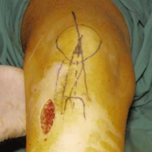
The patella may dislocate in a normal knee with an injury in which the knee turns in while the foot is stuck or turns out. The knee feels like it comes apart. The kneecap dislocates to the outer side of the knee (FIG. 1 Below) but often it appears that there is a prominent bone towards the inner side of the knee which is due to the way the knee is twisted.
If the kneecap stays out, the best solution is to try to relax the muscles (despite the pain), and have somebody help pull the knee out straight. The kneecap then drops back into place with instant relief of pain.
The kneecap may go back into place before you see it in the dislocated position which can make the diagnosis difficult afterwards.
Once the kneecap is back in place, the knee swells and becomes generally stiff and sore so the diagnosis may not be easy. However, careful examination should reveal tenderness at the inner edge of the kneecap and/or at the inner bony prominence of the knee (medial epicondyle), indicating tearing of the check-rein (medial retinaculum) that is meant to hold the kneecap in place and prevent excessive lateral displacement.
Damage to the joint surfaces of the kneecap and/or the outer edge of the knee (lateral femoral condyle) may result from the kneecap grating over the edge of the bone as it goes out and/or back into place. This may cause a feeling of grating or clicking in the knee afterwards and interfere with recovery.
Sometimes the kneecap does not completely dislocate. Subluxation is the term used to describe when the kneecap goes only partly out of place.

Recurrent Patella Dislocation
Sometimes further twisting episodes may result in further episodes of the kneecap dislocating. This may occur more and more easily and not result in all that much aggravation of pain and swelling afterwards. The knee becomes insecure with any twisting movement.
There are many predisposing anatomical factors which may contribute to recurrent patella instability including:
Shallow trochlea groove (the groove the knee-cap glides in)
Underdeveloped (dysplastic) patella
Poor muscle control from the lower, inner part of the quadriceps muscle (VMO – Vastus Medialis Obliquus). This muscle has to hold the knee cap in the groove as it tracks.
Laxity or detachment of the inner check reign of the kneecap (MedialPatello-Femoral Ligament: MPFL)
Tightness of the outer check rein (lateral retinaculum)
Excessive change of direction between the pull of the quadriceps tendon above and the patella tendon connecting the kneecap to the top of the shin (increased Q angle) FIG. 2
“Squinting Patellae”: turned-in knees with turned-out feet FIG.3
High riding kneecap (patella alta)


Recurrent Patella Subluxation
The kneecap may be unstable, causing a feeling of insecurity with twisting movements but without any episodes of actual dislocation. Often it is not possible for you to know what exactly happens at the time of these episodes or even that it is the kneecap that is the problem. This can make accurate assessment and diagnosis difficult, but there is a test called the Patella Apprehension Test which can be very helpful. The professional examining your knee tries to push the knee-cap outwards and if that reproduces the feeling that something may be about to go out of place, the diagnosis is confirmed.
If this test simply reproduces some pain, it may be that the insecurity is caused more by catching or pinching underneath the kneecap than through instability of the kneecap shifting out of place, or you may have a combination of both factors.
The same anatomic factors that predispose to recurrent patella dislocation also predispose to recurrent patella subluxation, and in fact, most people with recurrent patella dislocation also suffer many subluxation episodes between major dislocation episodes.
Patella Mal-Tracking
The knee-cap may fail to track centrally in the groove in a variety of ways and this may be very subtle, such that it is a relatively minor component of a combination of factors contributing to patello femoral pain. In that situation there is usually muscle weakness and perhaps wasting (thinning) of the lower inner part of the quadriceps (VMO), which allows the kneecap to glide a little off centre, predisposing to pain and further inhibition of muscle control. Sometimes this shows up on X-rays as a patella that is tilted, and/or shifted slightly off-centre. FIG.4
Another form of mal-tracking, associated with a tendency to recurrent patella subluxation is when the kneecap shifts outwards (laterally) as the knee straightens and has to shift inwards (medially) to re-enter the trochlea groove as the knee starts to bend again. This may feel like the kneecap jumps and may be uncomfortable. This particularly happens when the knee-cap sits high (Patella alta) and doesn't enter the groove until the knee is already bent some 30 degrees or so. Normally the patella should enter the groove and centre very soon after nee bending starts.
Another form of mal-tracking is due to excessive tightness and pressure on the outer half of the kneecap, but this usually causes pain rather than recurrent subluxation.This condition is called excess lateral pressure syndrome (ELPS). FIG.5


Treatment of Patella Instability
Quadriceps Rehabilitation
The key to successful treatment of patella instability is good quadriceps control of the tracking of the kneecap. Often this is achievable by a specific exercise program, which can be taught and supervised by a Physiotherapist. Physiotherapy is often required to help settle pain which may interfere with the exercises and to provide some temporary control of mal-tracking of the knee-cap to enhance the quadriceps ability to work and/or to control the tendency to recurrent subluxation episodes.
Whatever other treatment may be required, quadriceps rehab is the essential ingredient and once successful, a maintenance exercise program should be continued.
Avoidance of Risk Factors
Your Physiotherapist can also help you to learn to avoid the kind of twisting movement that provokes episodes of patella instability. This movement occurs when the knee twists inwards, especially if the foot is pointed outwards by comparison. Learning to avoid letting the knee twist inwards, trying always to keep the knee pointing in the same direction as the toes, is important if surgery is to be avoided. This may translate to feeling like you are running, and especially side stepping, on a wider base than you are used to.
Knee Support
Your Physiotherapist may tape the kneecap inwards to help stabilise it and help facilitate your quadriceps rehab. If that is effective, it may also be worth trying a patella stabiliser knee support which looks a little like a padded horse-shoe that sits around the outer half of the knee, held in place by three straps that wrap around the knee with Velcro fasteners.
Other simple knee supports may improve confidence without necessarily actually holding the kneecap in place.
Patella Realignment Surgery
a) Lateral Release
Sometimes, particularly if an arthroscopy is required after an acute dislocation episode has caused some joint surface damage, a lateral release of the patella alone may improve the imbalance, if the lateral retinaculum is tight.
b) Distal Patella Tendon Transfer
This operation involves detaching the piece of bone at the top of the shin (the tibial tuberosity) into which the patella tendon inserts and shifting it inwards about 1cm, to reduce the Q angle between the alignment of the patella tendon below, and the alignment of the quadriceps tendon above the kneecap. FIG. 6 This is often enough to rebalance the patella in the groove and enable quadriceps control to prevent recurrent dislocation episodes. This is a fairly small operation and the bone is fixed in place with a screw and the knee is protected with a removable straight knee splint until good muscle control is regained over the following 3 to 4 weeks.
Sometimes the patella is quite high-riding, so that the knee has to bend more than 30° before the patella enters the groove. In these cases it is a good idea to also bring the kneecap down to its correct height, by also “distallising” the tibial tuberosity, as well is shifting it inwards.


c) Medical Plication
The looseness of the medial patella retinaculum often needs to be tightened to really effectively stabilise the patella. Usually this is achievable through a small incision along the inner margin of the knee-cap. The loose retinaculum is divided and either a strip of tissue is excised, or the loose tissue is double breasted onto the kneecap to effectively tighten this structure. FIG. 8. This is often done in combination with a distal realignment and lateral release, but if done in isolation, it is not necessary to wear a knee splint afterwards. It nevertheless takes a week or two to regain quadriceps control and crutches are required for the first few days. Quads strength is regained after six to twelve weeks of rehabilitation. Sometimes, however, this plication can stretch out again with time, which is why there is now a growing trend to the next procedure.


d) Reconstruction of the Medial Patello-Femoral Ligament (MPFL)
The medial patello-femoral ligament (MPFL), which attaches from the bony prominence at the inner side of the knee (the medial epicondyle) to the inner edge of the kneecap, is the vital part of the medial retinaculum that prevents the patella from going out. If the MPFL is no longer attached to the medial epicondyle, simply tightening the retinaculum is not usually sufficient and the retinaculum subsequently works loose again. At the time of doing a medial plication, it is important to check that there is some attachment to the medial epicondyle, and if not, a graft can be used to reconstruct the medial patello-femoralligament. Sometimes a tendon such as semi-tendinosis, or the inner edge of the lower end of the quads tendon is used for that purpose, but the strip of loose retinaculum that is excised, can also be used for that purpose. FIG. 9
It is very important that this reconstruction not be made too tight, but when it is done correctly, especially in combination with the other realignment procedures described here, it can produce the most reliable results in terms of restoring patella stability. The recovery is no more involved following the reconstruction of the medial patellofemoral ligament than after a simple medial plication procedure (+/- the distal realignment of the patellatendon), although sometimes it can be a bit slower.
Conclusion
Every individual is different and it is important that your physiotherapist and your surgeon identify each aspect of the problem that is contributing to not only the instability and/or maltracking, but also any problems contributing to pain, which may undermine the success of your rehab.They will discuss with you what is to be done and give you some idea of the likely quality of the outcome, but there is no operation that can be guaranteed. The speed and quality of people’s recovery is as varied as the people themselves, and complications can and do occur from time to time, even though we take every precaution to minimise risks. If your recovery is a bit slow and bumpy, we will be there to help you through until the best possible outcome is achieved.